


Vitamin D Testing
From Nobel Discovery to Everyday Primary Care
By Dr Eric Law
Published September 2026
Abstract
Vitamin D occupies a distinctive position at the intersection of nutrition, photobiology and endocrinology. First identified in the early twentieth century as the antirachitic factor responsible for preventing rickets, it is now recognised as a secosteroid hormone exerting genomic effects through a nuclear receptor. Its scientific journey spans industrial public health crises, sterol chemistry, ultraviolet photochemistry, molecular endocrinology and more. For many of us, Vitamin D remains relevant in skeletal health, falls prevention and selected high-risk populations. Revisiting its discovery and biochemical evolution illustrates how a subtle ultravioletinduced modification of a cholesterol derivative continues to influence everyday clinical decision-making.
Introduction: A Disease of Industrialisation
Long before Vitamin D was recognised as a vitamin, its precursor molecules were absorbing ultraviolet radiation in primitive eukaryotic organisms inhabiting the ancient oceans. Ergosterol, a Vitamin D₂ precursor and fungal membrane sterol present in early phytoplankton, underwent photochemical transformation when exposed to sunlight, an evolutionary adaptation that may have provided protection against ultraviolet damage. Hundreds of millions of years later, a closely related photochemical principle would prove central to understanding one of the most devastating diseases of industrial childhood.
At the turn of the twentieth century, rickets was endemic in industrialised cities across Europe and North America. Rapid urbanisation had drawn families into densely populated, smoke-laden environments where narrow streets and coal-derived smog obscured direct sunlight. In some industrial centres, autopsy studies suggested that as many as 80–90% of children exhibited evidence of rickets. The skeletal manifestations were striking: bowed legs, widened wrists, rib deformities, growth retardation and profound muscular weakness. Pelvic deformities in affected girls later contributed to lifethreatening complications in childbirth. The link between industrialisation and skeletal deformity was unmistakable, yet the biological mechanism remained mysterious.
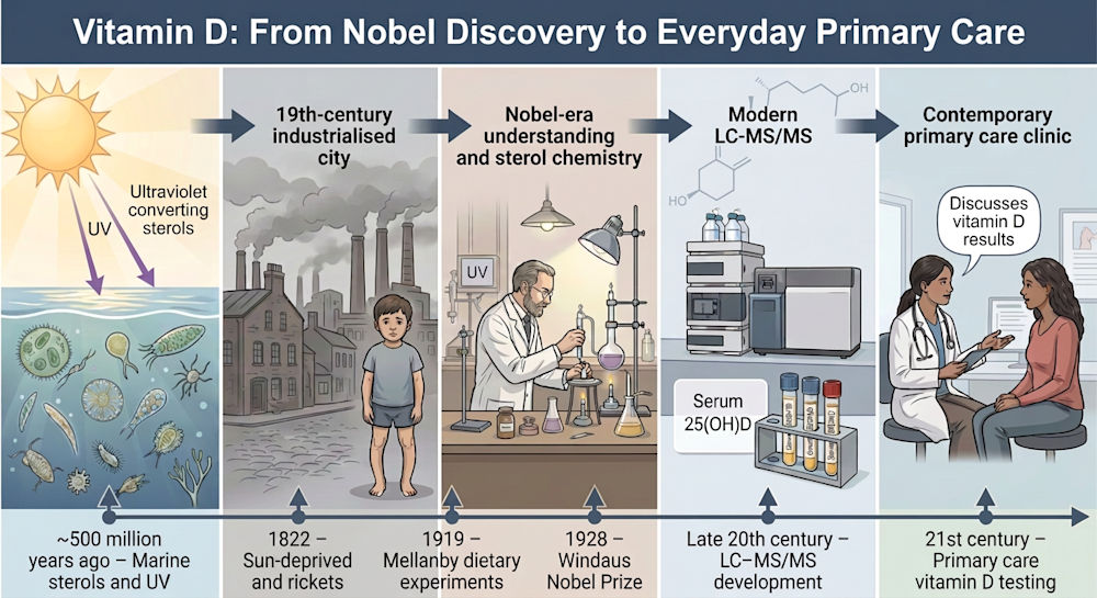
Clinical observation preceded biochemical explanation. In 1822, the Polish physician and
chemist Jędrzej Śniadecki noted that children in sun-deprived Warsaw developed rickets, whereas rural
children exposed to sunlight did not. Later, in 1890, Theobald A. Palm recognised the geographic
relationship between limited sunlight and the high incidence of rickets in urban Britain.
Collectively, these observations laid the groundwork for the discovery of the “sunshine vitamin”.
Meanwhile, fishing communities had long administered cod liver oil to prevent the disease, which was
later recognised as a rich source of Vitamin D. In 1919, Sir Edward Mellanby demonstrated
experimentally that rickets could be induced in porridge-fed, caged dogs through dietary restriction
and reversed with cod liver oil, implicating a nutritional deficiency. Professor Elmer McCollum, later
referred to by TIME magazine as “Dr Vitamin” for his discovery of Vitamin A in 1913 and Vitamin
B in 1915, subsequently established that the antirachitic factor was a new and distinct vitamin,
coined Vitamin D in 1922.
Ultraviolet radiation soon emerged as a critical component in elucidating the pathogenesis and treatment of rickets. Dr Kurt Huldschinsky demonstrated that exposure of affected children to mercury arc lamps resulted in radiographic mineralisation of long bones, thereby providing compelling evidence that ultraviolet irradiation induced a cutaneous photochemical reaction generating a circulating antirachitic factor. The subsequent structural elucidation of this factor was advanced through developments in sterol chemistry. Cholesterol, initially identified in human gallstones, was subsequenty and unexpectedly recognised as central to the biochemical framework of Vitamin D. Dr Adolf Windaus subsequently demonstrated that ultraviolet irradiation of a cholesterol precursor yielded the antirachitic compound, thereby establishing Vitamin D as a sterol-derived molecule activated by photochemical transformation. In recognition of his work elucidating the relationships among sterols, vitamins and bile acids, and determining their structures, Dr Windaus was awarded the Nobel Prize in Chemistry in 1928.
The Sterol Backbone: Structure Determines Function
What began as an investigation into a crippling disease of industrial childhood ultimately revealed a profound biological principle: sunlight can convert a membrane sterol into an endocrine signal. From ancient marine photochemistry to the smoke-darkened streets of industrial Europe, Vitamin D emerged as a molecule that links environment, molecular structure and hormonal regulation in a single elegant pathway. To understand Vitamin D further, one must begin with sterols.
Sterols are a subgroup of steroids and a class of lipids characterised by a four-ring carbon core, a hydroxyl (–OH) group at the C3 position and typically a side chain at C17.
All steroids share a common tetracyclic framework of four fused carbon rings (A, B, C and D), known as the cyclopentanoperhydrophenanthrene nucleus, the classical steroid backbone. Rings A, B and C are six-membered, while ring D is five-membered. This four-ring system is believed to enhance chemical stability and optimise function. Cholesterol is a sterol, whereas cortisol, estradiol, testosterone and aldosterone are steroid hormones derived from the same structural framework.
Vitamin D (D3) originates from 7-dehydrocholesterol, a cholesterol derivative synthesised in the skin. When exposed to ultraviolet B radiation (approximately 290–315nm wavelength), a photochemical reaction cleaves the bond between carbon atoms 9 and 10 in the B ring. This reaction opens the B ring. The resulting molecule is not a classical steroid but a secosteroid (from the Latin secare, “to cut”). The A, C and D rings remain intact, but the opened B ring transforms a rigid membrane sterol into a conformationally flexible signalling molecule capable of endocrine activity. A single ultraviolet-induced structural modification thus generates profound physiological consequences.
From Photochemical Product to Active Hormone: Metabolism, Molecular Forms and Measurement
Vitamin D₃ (cholecalciferol), synthesised in the skin, is biologically inert. It functions as a prohormone and must undergo sequential hydroxylation to become hormonally active.
The first hydroxylation occurs in the liver, mediated primarily by Vitamin D 25-hydroxylase (CYP2R1), producing 25-hydroxyvitamin D [25(OH)D]. This metabolite represents the principal circulating form and the usual laboratory marker used to assess Vitamin D status in clinical practice. Its half-life of approximately 2-3 weeks reflects its role as a systemic reservoir rather than an active hormone.
The second hydroxylation occurs predominantly in the kidney, where 25(OH)D is converted by 1α-hydroxylase (CYP27B1) into 1,25-dihydroxyvitamin D [1,25(OH)₂D] (calcitriol), the active hormone with a half-life of around 4-6 hours. This step is tightly regulated by parathyroid hormone (PTH), fibroblast growth factor 23 (FGF23), serum calcium and phosphate concentrations, thereby integrating Vitamin D metabolism into the classical endocrine feedback mechanisms that maintain mineral homeostasis.
Extrarenal expression of CYP27B1 in immune cells, the placenta and other tissues enables local (paracrine or autocrine) production of calcitriol, adding further physiological complexity. Catabolism occurs via 24-hydroxylase (CYP24A1), which inactivates both 25(OH)D and calcitriol. Pathogenic variants in CYP24A1 impair degradation and may lead to hypercalcaemia and hypercalciuria, a clinically relevant genetic disorder of Vitamin D metabolism (also known as infantile hypercalcemia type 1, an autosomal recessive condition). Understandably, detailed exploration of genetic bone diseases and inherited Vitamin D disorders would take us well beyond the intended scope of this article.
The link between sunlight, Vitamin D and bone health represents a distinctive physiological adaptation. Unlike other vitamins obtained primarily through diet, Vitamin D can be synthesised in the skin from a cholesterol precursor following ultraviolet B exposure and subsequently acts as a hormone regulating gene transcription. This light‑dependent mechanism provides an environmental cue that supports calcium absorption and skeletal mineralisation, processes essential for maintaining structural integrity in terrestrial life. The uniqueness of this photochemical–endocrine pathway explains why Vitamin D, unlike other vitamins, plays such a central and irreplaceable role in bone health.
Molecular Diversity: D₂, D₃ and Epimers
Vitamin D exists in multiple biochemical forms reflecting its metabolic progression. These include Vitamin D₃ (cholecalciferol), derived from animal sources or by conversion of 7-dehydrocholesterol in the skin, and Vitamin D₂ (ergocalciferol), derived from plant and fungal sources.
Both forms undergo identical hepatic and renal activation pathways. Although structurally similar, D₂ and D₃ differ slightly in their side-chain configuration (D2 includes an additional double bond between C22 and C23 and a methyl group at C24), which may influence pharmacokinetics.
Metabolism also produces epimeric variants, most notably C3 epimers such as 3-epi-25(OH)D. Epimers are stereoisomers differing in configuration at a single chiral carbon—that is, carbon 3 of the A ring, where the usual hydroxyl group is flipped “downward” instead of “upward”. While structurally similar to 25(OH)D, the subtle but biologically significant spatial orientation of the hydroxyl group can reduce receptor affinity (e.g. the geometry of the hydrogen bond) and biological potency. Of note, C3 epimers can be found in neonates and infants; in adults, their clinical significance appears limited but analytically relevant.
Analytical Considerations
Clinical assessment measures serum 25(OH)D (unit: nmol/L), not 1,25-dihydroxyvitamin D (for which concentrations are typically measured in pg/mL rather than ng/mL). This distinction is crucial: 25(OH)D reflects cumulative Vitamin D stores, whereas 1,25(OH)₂D is tightly regulated and often normal even in deficiency.
Most routine immunoassays measure total 25(OH) D but may not reliably distinguish between D₂, D₃ and epimers. Cross-reactivity can modestly influence results, particularly in paediatric populations. Liquid chromatography tandem mass spectrometry (LC–MS/MS), when appropriately configured, can differentiate 25(OH)D₂, 25(OH)D₃ and epimers with greater specificity.
In addition, routine immunoassays may not fully distinguish the C3‑epimer from standard 25(OH)D. In most healthy adults, this analytical limitation has minimal clinical impact. However, it may become relevant in neonatal assessment, rare disorders of Vitamin D metabolism, research settings or when laboratory results appear discordant with the clinical picture. Where clinically indicated, specialised LC‑MS/MS testing with separation of individual Vitamin D species can be arranged. Please discuss this with a pathologist or relevant specialist if further clarification is required.
Genomic Signaling: The Vitamin D Receptor
Calcitriol exerts its biological effects by binding to the Vitamin D receptor (VDR), a ligand-activated transcription factor belonging to the nuclear receptor superfamily, which also includes receptors for glucocorticoids, mineralocorticoids, oestrogens, progesterone and thyroid hormone. In brief, nuclear receptors are specialised proteins inside cells that bind to small molecules such as hormones and act as transcription factors to “switch on” or “switch off” the genes.
Beyond the classical role in calcium and bone homeostasis, 1,25-dihydroxyvitamin D regulates the expression of numerous genes across a wide range of tissues, including immune cells, skeletal muscle, the vasculature and reproductive organs. Upon ligand binding, the VDR forms a heterodimer with the retinoid X receptor (RXR) and interacts with Vitamin D response elements (VDREs) in the promoter regions of target genes. This complex recruits coactivator or corepressor proteins, leading to chromatin remodelling and regulation of gene transcription. At the time of writing, the exact number of genes targeted or influenced by Vitamin D remains unknown. Estimates suggest it may be in the hundreds to over a thousand. Some examples include:
- Intestinal calcium and phosphate absorption
- Bone remodelling and mineralisation
- Suppression of parathyroid hormone
- Renal tubular calcium handling
- Cellular proliferation and differentiation
Vitamin D Testing in Practice: From Research to Clinical Laboratory
Given the central role of Vitamin D in calcium homeostasis, bone metabolism and its broader effects on immune, muscular and cardiovascular function, disturbances in Vitamin D status can have important clinical consequences. Both deficiency and excess may impact skeletal health and contribute to a range of systemic conditions. Therefore, modern guidance recommends selective, risk-based testing rather than routine population screening for Vitamin D status. Measurement of serum 25-hydroxyvitamin D [25(OH)D] is considered appropriate in individuals with specific risk factors or clinical indications. More information is available in standard guidelines and professional position statements.
A serum 25(OH)D threshold of 50 nmol/L is commonly used to define adequacy for bone health, particularly at the end of winter, in recognition of seasonal variation. According to the Royal College of Pathologists of Australasia (RCPA; Vitamin D – Interpretation of Results, accessed 22 July 2026), the following categories apply:
- Vitamin D adequacy: ≥50 nmol/L at the end of winter (levels may need to be 10-20 nmol/L higher at the end of summer to account for seasonal decline)
- Mild deficiency: 30-49 nmol/L
- Moderate deficiency: 12.5-29 nmol/L
- Severe deficiency: <12.5 nmol/L
Vitamin D toxicity is characterised by hypercalcaemia, hypercalciuria and nephrocalcinosis. Hypercalcaemia is uncommon until serum 25(OH)D concentrations exceed approximately 220 nmol/L and is more typically reported at levels above 500 nmol/L.
If laboratory results are discordant with the clinical presentation, or fail to respond appropriately to supplementation, measurement by LC-MS/MS may be considered for more specific assessment.
Conclusion: A Molecule Illuminated by Sunlight
Vitamin D reminds us that even the most familiar terms in everyday conversation may reflect intricate and elegant physiology, and that much of modern clinical practice remains grounded in chemistry, quite literally illuminated by light. Nevertheless, in routine practice, this remarkable cascade of photochemistry and endocrine regulation is ultimately distilled into a single numerical value (nmol/L) on an otherwise unremarkable laboratory report.
The modest figure, while clinically practical, signifies far more than a threshold to correct or a target to attain. It represents a finely regulated biological system shaped by evolution, environment and human behaviour. To recall the science behind the number is to restore perspective: even the most routine test result reflects molecular precision, physiological balance and the enduring interplay between sunlight and human biology.
How to Order Vitamin D Testing
Note the reason for testing to meet Medicare eligibility criteria for bulk-billing in the ‘Clinical Notes’ section on a Clinical Labs General Pathology Request Form.
Blood samples can be collected at any Clinical Labs collection centre.
Due to the long half-life, repeat testing should not be performed earlier than 3 months after either starting or changing the dose of vitamin D supplements.
Serum calcium, phosphate and parathyroid hormone will assist in placing the vitamin D level within the context of overall calcium homeostasis.
If osteoporosis is present, assessment of bone turnover markers (fasting C-terminal telopeptide of type 1 collagen [CTX] and procollagen type 1 N propeptide [P1NP]) may be considered to provide a way of monitoring bone turnover in response to therapy.
References
Holick MF. The One-Hundred-Year Anniversary of the Discovery of the Sunshine Vitamin D₃: Historical, Personal Experience and Evidence-Based Perspectives. Nutrients. 2023;15(3):593. doi:10.3390/nu15030593.
Norman AW. From vitamin D to hormone D: fundamentals of the vitamin D endocrine system. Am J Clin Nutr. 2008;88(Suppl):491S–499S.
Nowson CA, et al. Vitamin D in Australia: issues and recommendations. Aust Fam Physician. 2004;33(3):133–138.
Raczyk M, Carlberg C. From Sunlight to Signaling: Evolutionary Integration of Vitamin D and Sterol Metabolism. Metabolites. 2026;16(1):74. doi:10.3390/metabo16010074.
Royal Australian College of General Practitioners. First Do No Harm: Vitamin D testing. November 2022.
Royal College of Pathologists of Australasia (RCPA). Vitamin D (25 hydroxyvitamin D). RCPA Manual. Last reviewed 2 January 2024.
Royal College of Pathologists of Australasia. Use and Interpretation of Vitamin D Testing. Position Statement 1/2013. Updated December 2023.
Stott VE, Stilgoe S, Stroud ML. Vitamin D: A review. Aust Fam Physician. 2008;37(12):1002–1005.
The Lancet Diabetes & Endocrinology. Vitamin D—controversies and consensus in testing and supplementation. Lancet Diabetes Endocrinol. 2023. doi:10.1016/S2213-8587(23)00060-8.
About the Author

Dr Eric Law
MBChB BMedSc(1st Hon) PhD FHKAM(Pathology) FHKCPath FRCPA FRCPath FAACB FFSC(Research) IFCAPDr Eric Law
MBChB BMedSc(1st Hon) PhD FHKAM(Pathology) FHKCPath FRCPA FRCPath FAACB FFSC(Research) IFCAP- biochemical genetics
- chemical pathology
- inherited metabolic disorders
Dr Law completed his medical science degree (first honor) and MB ChB at the Chinese University of Hong Kong and obtained his PhD from the University of Hong Kong. He is a Fellow of the Royal College of Pathologists of Australasia (FRCPA), with scope of practice in both Chemical Pathology and Genetic Pathology. Additionally, he is a Fellow of the Australian Association of Clinical Biochemists (FAACB), the Royal College of Pathologists (FRCPath), an International Fellow of College of American Pathologists (IFCAP), Hong Kong College of Pathologists (FHKCPath), Hong Kong Academy of Medicine (FHKAM(Pathology)) and the Faulty of Science (Research), RCPA. His areas of interest include chemical pathology, inherited metabolic disorders, clinical metabolomics and medical teaching.
Local Pathologists
 Pathologist

 Pathologist
Related Pages

Thyroid Function Test Interpretation
Dr Phoebe Stanford explains how to interpret thyroid function tests accurately, addressing common pitfalls and interferences to improve diagnostic confidence.

Urine Testing in Asymptomatic Patients
From 1 July 2025, the Medicare Benefits Schedule (MBS) item for urine testing (69333) will be amended.

Assessing Allergy in Clinical Practice
Associate Professor Louise Smyth explains the clinical significance of allergy, immune responses and the role of accurate testing in managing allergic conditions.